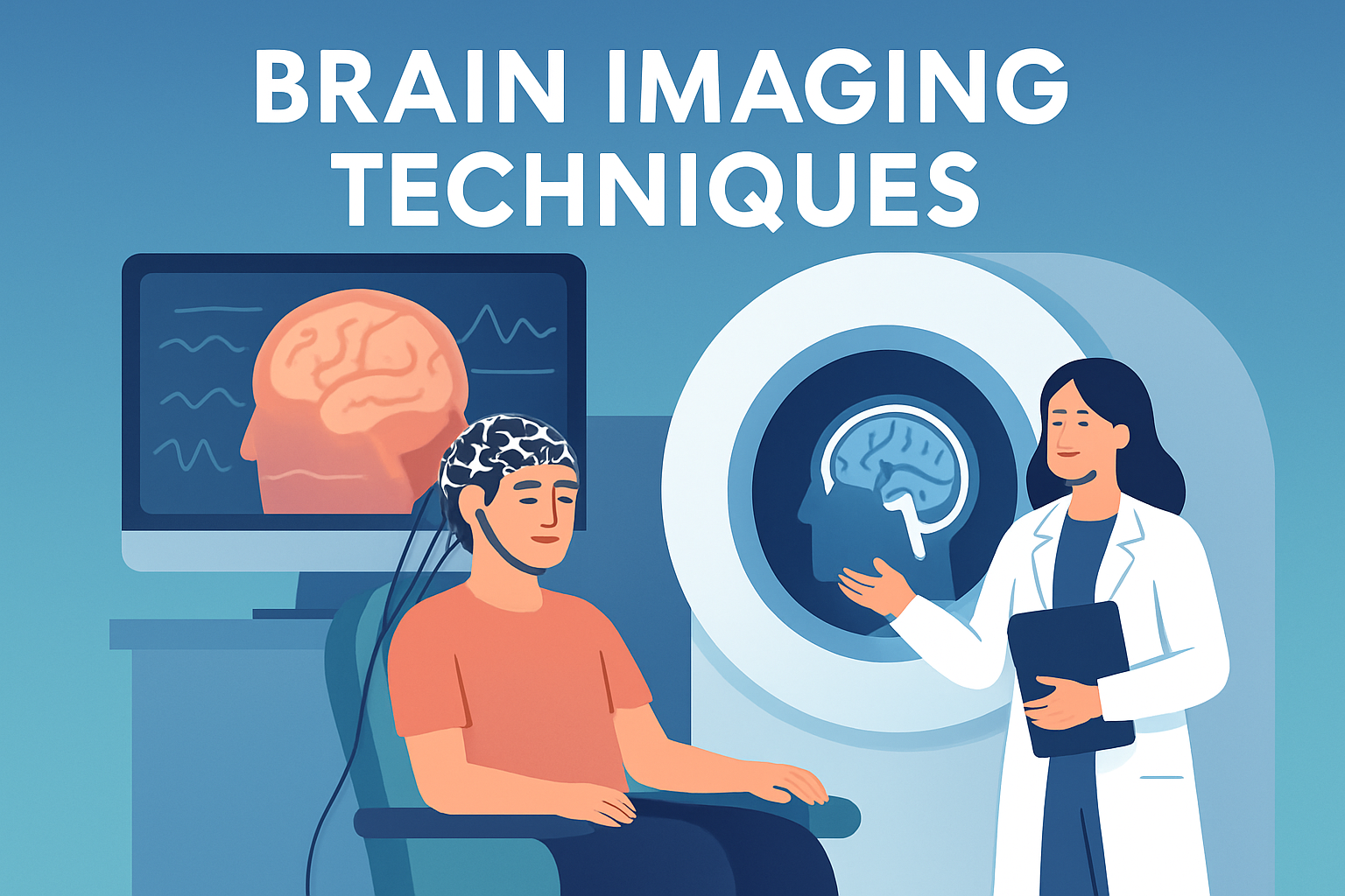
Understanding the human brain has long been a fascination for scientists, researchers, and medical professionals. As the command center of the human body, the brain’s functionality is complex and sophisticated. To study and comprehend its mechanisms, brain imaging has become an invaluable tool. It allows us to see inside the brain in a non-invasive way to observe structures and functions. Among the most widely used techniques are fMRI (functional Magnetic Resonance Imaging), PET (Positron Emission Tomography), and EEG (Electroencephalography). These techniques have revolutionized neuroscience, allowing breakthroughs in understanding neurological diseases, human cognition, and brain functions. Each method has its unique advantages, applications, and limitations, making them suitable for different types of research and clinical applications.
The importance of these brain imaging techniques cannot be overstated. They play key roles in diagnosing and treating conditions like Alzheimer’s disease, epilepsy, and schizophrenia. Besides, they are crucial in the field of cognitive neuroscience, contributing to our understanding of brain behavior relations, decision-making processes, and even emotions. Moreover, these imaging techniques have broadened their impact beyond healthcare, influencing fields such as psychology, where they aid in better exploring human consciousness and behavioral studies, making it a significant area of research and application.
Functional Magnetic Resonance Imaging (fMRI)
Functional Magnetic Resonance Imaging, commonly known as fMRI, primarily measures brain activity by detecting changes associated with blood flow. This technique utilizes magnetic fields and radio waves to create detailed images of the brain. When a brain area is in use, blood flow to that region also increases, which can be measured and mapped using fMRI, providing both functional and structural information. This is hugely beneficial for understanding which areas of the brain are involved in particular mental processes.
A real-world example of fMRI in action is in language studies. Researchers can use fMRI to identify brain areas activated during language processing tasks. For example, when a person is asked to perform a language task, such as listening to a story, specific brain regions involved in language processing, like Broca’s and Wernicke’s areas, show increased activity. This helps researchers understand language disorders and develop targeted therapies.
One of the main advantages of fMRI is its high spatial resolution, enabling scientists to pinpoint precise brain areas involved in specific tasks. However, a downside is its relatively low temporal resolution. It can’t capture rapid neuronal activity because it measures blood flow changes, which lag behind neuronal firing by a few seconds. Despite this, the wealth of information provided by fMRI makes it invaluable in current neuroscience research.
Positron Emission Tomography (PET)
Positron Emission Tomography, or PET, is another powerful imaging tool used to observe brain processes. Unlike fMRI, PET scans involve the injection of radiotracers into the bloodstream. These tracers are often attached to biologically active molecules like glucose, allowing one to visualize brain metabolism. As the brain consumes these tracers, a PET scanner detects the emitted positrons, generating detailed images reflecting metabolic activity.
A significant application of PET is in oncology, detecting early signs of cancer by observing metabolic changes before structural ones become evident. In neurological applications, PET scans are used to diagnose and monitor diseases such as Alzheimer’s by visualizing amyloid plaques accumulation. By receiving a radioactive tracer that binds to these plaques, PET can accurately track disease progression over time.
Despite its usefulness, PET has limitations. Its resolution is not as high as fMRI, and the use of radioactive materials, although safe, presents a certain level of risk, limiting its use primarily to clinical settings where its benefits outweigh potential downsides. PET scans are quite costly, and the preparation of specific radiotracers requires sophisticated facilities.
Electroencephalography (EEG)
Electroencephalography, abbreviated as EEG, is an imaging technique designed to capture electrical activity in the brain. By placing electrodes on the scalp, EEG records the electrical signals produced by the brain’s neuronal activity. This technique is particularly helpful for diagnosing conditions like epilepsy and sleep disorders.
A practical example of EEG use is in the diagnosis of epilepsy. An EEG can detect abnormal patterns signaling seizures, enabling healthcare professionals to determine the type of epilepsy and appropriate treatment. EEG is also used in sleep studies to track the sleep stages and diagnose disorders such as insomnia and sleep apnea.
One of the significant advantages of EEG is its superior temporal resolution, allowing it to capture rapid neuronal events and changes in real-time. However, it lacks the spatial resolution of fMRI and is less effective in pinpointing exact areas of activity. EEG signals can also be affected by artifacts from external electronic devices and even the person’s movements, necessitating a controlled environment for accuracy.
Comparison of Techniques
| Technique | Type of Imaging | Main Use | Advantages | Limitations |
|---|---|---|---|---|
| fMRI | Functional | Mapping brain activity | High spatial resolution | Low temporal resolution |
| PET | Functional/Metabolic | Measuring metabolic processes | Visualizes metabolic activity | Involves radioactivity |
| EEG | Functional | Capturing electrical activity | High temporal resolution | Low spatial resolution |
The variety in brain imaging techniques reflects the diverse needs of neuroscientific research and clinical diagnostics. Each method offers a unique lens into understanding the brain’s function and structure, making them complementary rather than competitors in the field of neuroscience.
Conclusion
In summary, understanding the capabilities and limitations of fMRI, PET, and EEG is crucial for selecting the appropriate technique for specific research or clinical purposes. fMRI offers detailed spatial insights into brain activity, beneficial for cognitive studies and mapping brain functions. PET provides valuable metabolic information, especially crucial in diagnosing illnesses like Alzheimer’s and cancer, while EEG shines in recording electrical activity, ideal for conditions like epilepsy and sleep disorders.
The continued advancement of these technologies promises ongoing breakthroughs in neuroscience and healthcare. The ever-expanding capabilities of imaging technologies continue to transform how researchers explore brain activity and diagnose neurological conditions. For practitioners and researchers, staying informed about the latest advancements and applications of these techniques is vital. We encourage readers involved or interested in this field to explore educational opportunities, workshops, or journals focused on these imaging techniques to stay abreast of the latest developments.
As technology advances, so too will our capacity to understand and heal the human brain, driven by the information and insights offered through these powerful imaging techniques.
If you’re interested in learning more, consider seeking courses that delve deeper into neuroimaging or engaging with professional organizations that focus on brain imaging research. The opportunities for deeper understanding and application are vast and continually expanding, promising a future rich with discovery and innovation in neuroscience.
Frequently Asked Questions
1. What is fMRI and how does it work?
Functional Magnetic Resonance Imaging (fMRI) is a brain imaging technique that measures and maps brain activity. It works by detecting changes in blood flow and oxygen levels in the brain. When a particular area of the brain is more active, it consumes more oxygen; blood flow to that region increases to meet the demand. The fMRI detects these changes in blood oxygenation and flow, creating detailed images, often in real-time, of brain activity. This technique does not use radiation, making it safer for repeated use to observe brain functions during different tasks or rest periods. It is particularly useful for understanding the role of specific brain regions in cognitive processes, emotional responses, and sensory tasks.
2. How does PET imaging differ from fMRI?
Positron Emission Tomography (PET) imaging is another widely used brain imaging technique that provides unique insights compared to fMRI. While fMRI measures changes in blood flow related to brain activity, PET works by detecting gamma rays emitted by a radioactive tracer that is injected into the bloodstream. This tracer, often linked to glucose, highlights areas of the brain with higher metabolic activity, offering insights into both the structure and the biochemical processes ongoing in the brain. PET is particularly useful for examining brain diseases, detecting cancerous cells, and analyzing brain function in conditions like epilepsy or dementia. The main difference between PET and fMRI is that PET can show more detailed information about biochemical processes and is sometimes used in conjunction with fMRI to provide a more comprehensive view.
3. What is EEG and how is it used in brain imaging?
Electroencephalography (EEG) is another essential technique in brain imaging, distinct from fMRI and PET because it is used to measure electrical activity in the brain. It involves placing electrodes on the scalp to detect electrical signals produced by the neurons. EEG is excellent for tracking brain activity over milliseconds, which is crucial for understanding rapid brain responses. It is often employed to diagnose conditions related to abnormal electrical brain activity, such as epilepsy, sleep disorders, and brain diseases. Although its spatial resolution is less detailed compared to fMRI and PET, EEG offers superior temporal resolution, allowing scientists and clinicians to track fast brain processes in real time.
4. What are the advantages of using fMRI, PET, and EEG in brain studies?
The array of brain imaging techniques—fMRI, PET, and EEG—each have distinct advantages, making them invaluable tools for research and clinical practice. fMRI offers exceptional spatial resolution, allowing for detailed mapping of brain structures and activities. It is non-invasive and does not involve radiation, making it ideal for longitudinal studies. PET provides valuable insight into brain metabolism and chemical processes, proving useful in detecting diseases like Alzheimer’s by identifying changes in cellular activity. Lastly, EEG’s rapid measurement of electrical activity is unparalleled, presenting an excellent choice for studying brain dynamics and neurophysiological responses. Together, these methods complement each other by providing a multifaceted view of brain structure and function.
5. Are there any risks or limitations associated with brain imaging techniques like fMRI, PET, and EEG?
While brain imaging techniques have revolutionized our ability to study the brain, they do come with some limitations and risks. fMRI, for instance, requires participants to remain still, which can be a challenge for some, especially young children or people with claustrophobia. Moreover, while it provides exquisite images, it cannot distinguish between different types of brain tissue as clearly as some other imaging methods. PET involves the use of radioactive tracers, so exposure must be limited, although the actual radiation dose is relatively low. This makes it less suitable for repeated measurements in quick succession or in certain populations, such as pregnant women. EEG is safe and non-invasive, but it does not provide high spatial resolution images, limiting the ability to pinpoint exact brain regions involved in specific tasks or responses. Nevertheless, balancing these limitations, the benefits of these imaging techniques in advancing our understanding of the brain are substantial.